
Fig. 4.
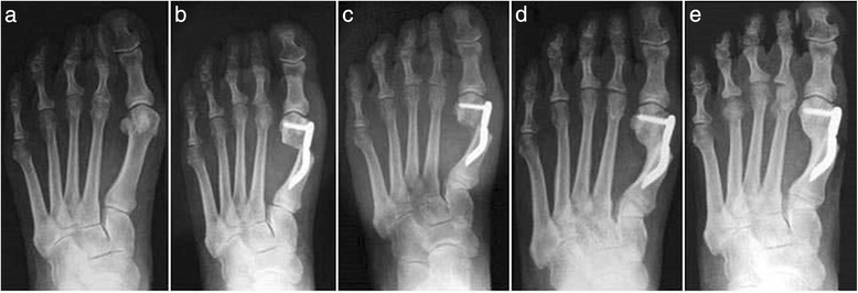
Case 1. A 44-year-old woman with mild hallux valgus (HV). a Preoperative antero-posterior radiographic image. b Endolog translation more than 100 % at 1-month follow-up, X-ray image. c Bone callus formation at 3-month follow-up, X-ray image. d a at 6-month follow-up X-ray image. e Radiographic aspect at 48-month follow-up showing the maintained correction of the deformity and complete healing of the osteotomy