
Fig. 3.
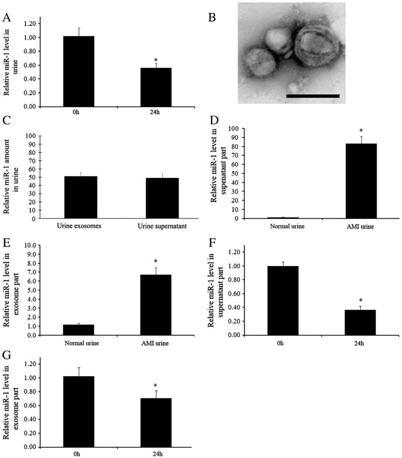
Stability and distribution of urine miR-1 in rats with AMI. (A) Urine samples were collected from rats at 24 h after AMI and miR-1 levels were determined immediately after urine collection and 24 h later at 4 °C. n=5, *P<0.01 compared with 0 h group. (B) Representative urine exosomes isolated by ultracentrifugation and detected by electron microscopy. (C) The distribution of urine miR-1 in exosome part and supernatant part (100% of total urine miR-1). (D) miR-1 levels in supernatant part of urine in rats with and without AMI. (E) miR-1 levels in exosome part of urine in rats with and without AMI. (F) miR-1 levels in freshly isolated supernatant part of AMI urine and in supernatant part kept for 24 h at 4 °C. (G) miR-1 levels in freshly isolated exosome part of AMI urine and in exosome part kept for 24 h at 4 °C. Note: n=5, *P<0.01 compared with 0 h group.