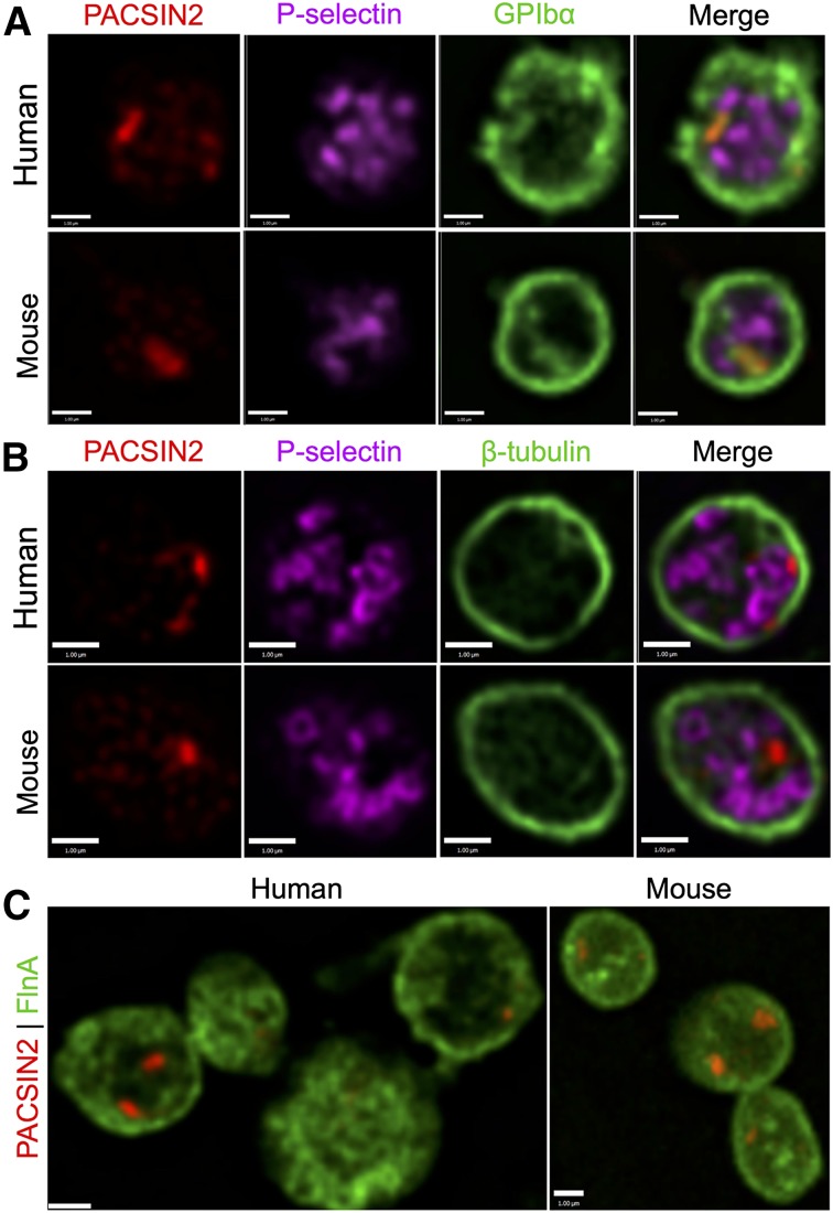
Figure 4.
PACSIN2 localization in human and mouse platelets. High-resolution SDM of (A) PACSIN2 (red), P-selectin (magenta), and GPIbα (green) in fixed human and mouse platelets; (B) PACSIN2 (red), P-selectin (magenta), and β-tubulin (green) in fixed human and mouse platelets; and (C) PACSIN2 (red) and FlnA (green) in fixed human and mouse platelets. Most platelets show 1 to 2 distinct concentrated PACSIN2 foci, which may also be distributed throughout the cells. Scale bars represent 1 µm.

