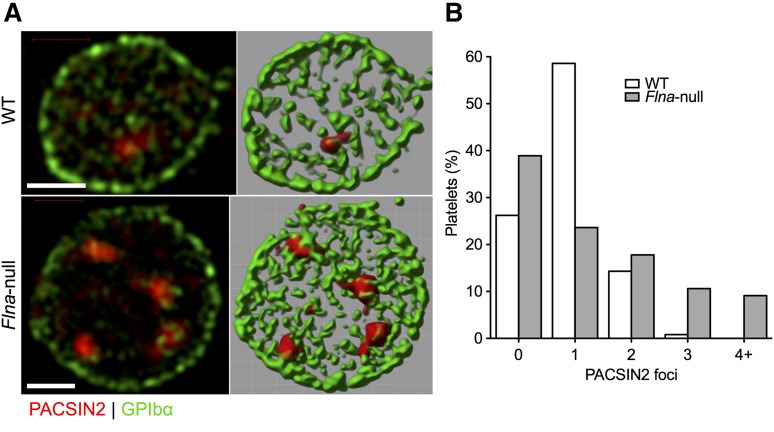
Figure 5.
PACSIN2 localization in WT and Flna-null platelets. (A) Super-resolution SIM of PACSIN2 (red) and GPIbα (green) in typical fixed WT and Flna-null mouse platelets (left). Fields were rendered in Volocity extended focus mode (right). Scale bars represent 1 µm. (B) Fields rendered were scored for the presence of defined PACSIN2 foci at a constant contrast setting. Total platelets scored were 244 WT and 208 Flna null.

