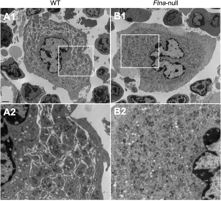
Figure 7.
Altered ultrastructure of Flna-null bone marrow MKs. Transmission electron microscopy analysis of freshly isolated bone marrow (A) WT and (B) Flna-null MKs. Areas within boxes in micrographs A1 and B1 are shown at higher magnification in A2 and B2, respectively. Scale bars represent 2 µm at ×5000 magnification (upper) and 500 nm at ×15000 magnification (lower).

