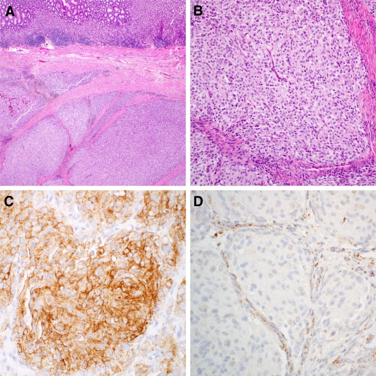
Figure 1.
Micrographs of a succinate dehydrogenase (SDH)-deficient gastrointestinal stromal tumor (GIST). Hematoxylin and eosin staining demonstrates typical multinodularity and intervening fibrous septae ([A]: low power; [B]: medium power), KIT immunohistochemistry shows strong KIT expression (C), and SDHB immunohistochemistry shows loss of SDHB expression in the GIST cells, with retained expression in the (noncancerous) fibrous septae as a positive internal control (D).

