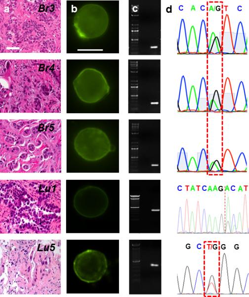
Figure 4. Single cell genomics of CTCs from patients.

(a) H & E staining of the primary tumor of metastatic breast and lung cancer patients. Tissue biopsies were used to determine the presence of DNA mutations on the oncogene PIK3CA and EGFR. (b) Panel of CTCs from the same metastatic breast and lung cancer patients in (a). Micrographs of the CTCs identified and subsequently released for molecular analysis using our selective release mechanism (scale bar 10 μm). (c) Micrographs of amplified DNA of the single CTCs shown in (b). (d) Sequencing of the amplified DNA from the single CTCs shown in (b). The 3140A/G (H1047R) point mutation in the PIK3CA oncogene as well as the exon 19 deletion and the 2573T/G (L858R) point mutation in the EGFR oncogene were detected at the single cell level.
