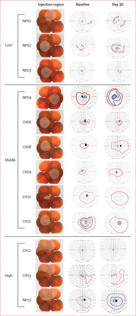
Figure 1. Area of retina exposed to adeno-associated virus-mediated delivery of wild-type retinal pigment epithelium (AAV2-hRPE65v2).
Column 1 was drawn over composite photographs of a normal retina, and columns 2 and 3 over the baseline and follow-up Goldmann visual fields, respectively, in the injected eyes. All follow-up visual fields are shown at day 30, except for patients NP01 (4·75 months) and NP02 (2·75 months). Stimuli used to measure Goldmann visual fields were V4e (red) and II4e (blue). Scotomas and the natural blind spot are shown in black. *Visual field data from these patients were reported previously1 but are presented here for completeness.

