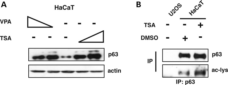
Figure 1.
The ΔNp63α protein is acetylated in human keratynocytes. (A) Western Blot (WB) analysis of whole HaCaT cell extracts treated with increasing amounts of TSA (5 ng/ml and 10 ng/ml) for 5 h or VPA (0.5 mm and 1 mm) for 3 h. (B) Whole cell extracts from HaCaT cells treated with 5 ng/ml of Trichostatin (TSA) for 5 h were analyzed by immunoprecipitation of endogenous ΔNp63α with an anti-p63 antibody followed by WB analysis with an anti-acetylated lysines. U2OS cells, not expressing p63, were used as negative control.

