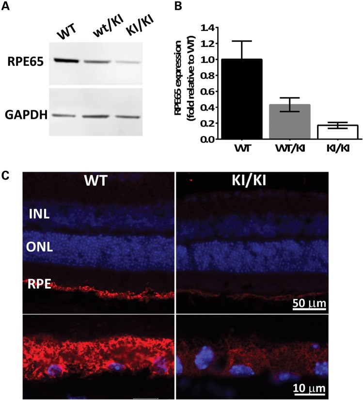
Figure 2.
Reduced mutant protein levels in the KI/KI mice. (A) RPE65 protein expression in eyecups was determined by western blot analysis in WT, heterozygous wt/KI and homozygous KI/KI (KI) mice. GAPDH protein expression was used as loading control. Representative blot shown; (B) quantification of RPE65 expression after normalization with GAPDH. Error bars indicate SDs from biological triplicates within the experiment; (C) immunofluorescence of eyeball sections from WT and KI mice with antibody against RPE65 (red), and nuclear staining with 4′,6-diamidino-2-phenylindole (DAPI; blue). The upper panels cover the full span of the retina, whereas the lower panels are higher magnifications of the RPE. INL, inner nuclear layer; ONL, outer nuclear layer; RPE, retinal pigment epithelium.

