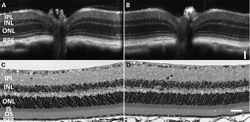
Figure 3.
Retinal histology of Rpe65P25L/P25L KI mice is similar to WT. Retinal integrity at the age of 7 months was monitored by both in vivo OCT imaging (A and B) and toluidine staining of the eyeball sections (C and D). There are no detectable differences in retinal structure between KI/KI (B and D) and its WT siblings (A and C) when raised under normal dim light conditions. Scale bar = 50 µm.

