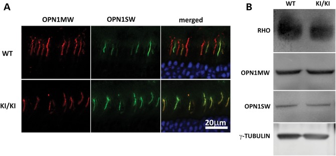
Figure 4.

Expression and localization of opsins. (A) Retinal sections of WT (upper) and Rpe65P25L/P25L (lower) mice were stained with antibodies against short-wave-sensitive opsin 1 (OPN1SW) and medium-wave-sensitive opsin 1 (OPN1MW). Both were correctly localized to the OS in the KI/KI mice. (B) Western blotting of opsin proteins from 7-month-old WT or Rpe65P25L/P25L mice. Similar levels of rod (RHO) or cone opsins were detected in retinae of mutant mice as in the WT mice.
