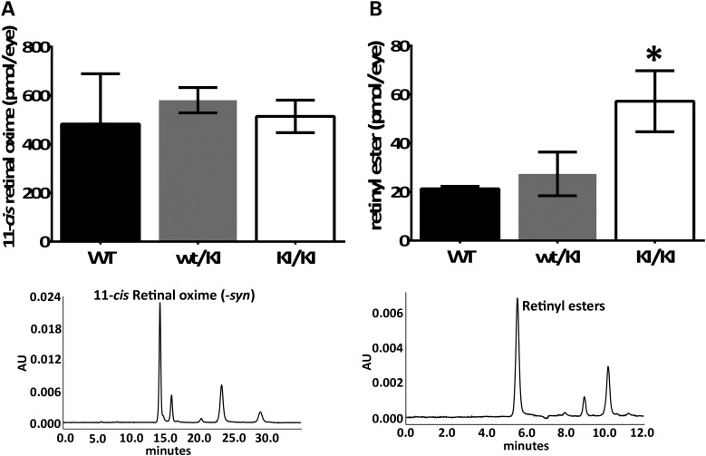
Figure 5.
Analysis of visual cycle retinoids from Rpe65P25L/P25L KI mice at the age of 4 months. (A) Quantitation of HPLC peak (lower panel, representative example of chromatogram) of 11-cis-retinyloxime from dissected retinae shows comparable amount of 11-cis-retinal present in the KI/KI mice as in the WT controls. (B) Quantitation of HPLC peak (lower panel, representative example of chromatogram) of total retinyl esters extracted from dissected eyecup samples showed ester accumulation in RPE. Error bars indicate SDs from biological triplicates within the experiment; *P<0.05.

