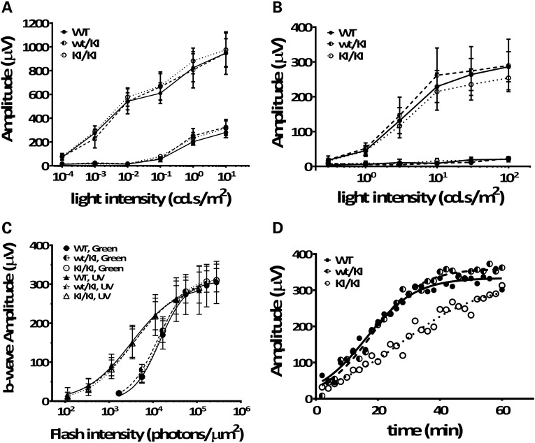
Figure 6.
Rpe65P25L/P25L KI mouse has full-field scotopic (rod), photopic (cone) ERG responses similar to WT but has delayed a-wave recovery after moderate visual pigment bleach. Dark-adapted (scotopic) ERG (A) and light-adapted (photopic) ERG (B) b-wave responses were obtained from 3-month-old homozygous KI/KI (open circle), heterozygous wt/KI (half-filled circle) and the littermate WT (filled circle) controls. Each point represents the average of six mice. Error bars show ±SD. (C) Cone signal isolation in Rpe65P25L/P25L KI mouse. B-wave amplitudes in response to a series of flashes varying intensity of UV (triangles) and green light (circles) stimuli were measured for 6-month-old homozygous KI/KI mice, heterozygous wt/KI and the littermate WT controls. Each point represents the average of four mice. Error bars show ±SD. (D) Representative a-wave recovery after moderate visual pigment bleach in KI/KI (open circle), WT (filled circle) and wt/KI (half-filled circle) mice. Continuous curves are plotted from Equation (1) fitted to the data with a nonlinear regression.

