Figure 7.
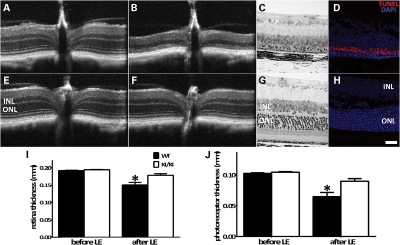
Rpe65P25L/P25L KI mice are protected from light damage. Eyeballs from WT (A–D) and KI/KI (E–H) mice were treated with high-intensity light (20 000 lux) for 30 min following pupil dilation. Retinal integrity was monitored by OCT before (A and E) and 2 weeks after light damage (B and F). Fine retinal structures after light exposure were examined by light microscopic analysis of sections of retinal tissues of WT mice (C) and KI/KI mice (G). Cell apoptosis after light treatment was surveyed via TUNEL staining of frozen sections of retinae of WT mice (D) and KI/KI mice (H). Thickness of the total retina (I) and the photoreceptor layer (J) before and after light damage was measured on 10 WT and 15 KI/KI mice. Error bars show ±SD; *P < 0.05.

