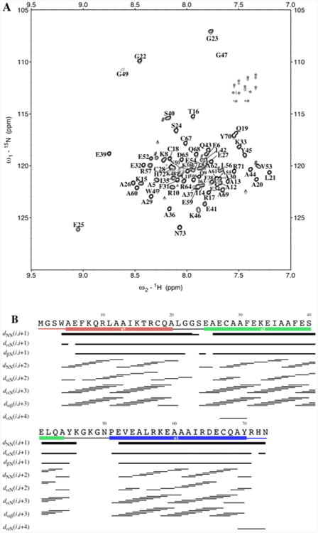
Figure 1.

(A) 15N-TROSY spectrum of 15N-labeled α3DIV. The assignments are adjacent to their corresponding peaks. Of the 73 residues, 69 were assigned, with residues 1–3 and Pro51 not observed in the spectrum. The residual Gln and Asn side-chain peaks were not assigned and are marked with an asterisk. Further, aliased and noise peaks are respectively given a pound and circumflex symbol. (B) Summary of sequential NOEs for apo α3DIV, which were determined from 3D 15N- NOESY-TROSY and 13C-NOESY-HSQC spectra.
