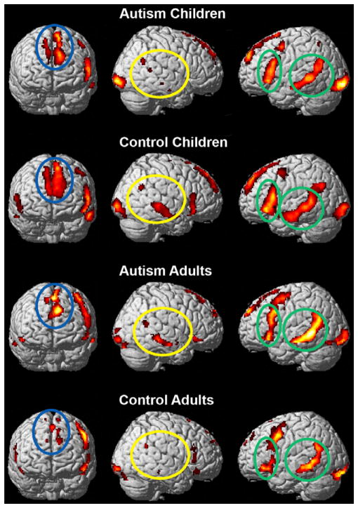
Figure 3.
Within-group brain activation in the irony versus fixation condition. The language-processing areas are more left lateralized for the children with autism and the adult controls. Figures are thresholded at P < 0.001, uncorrected. The green ellipses indicate left hemisphere language areas, the blue ellipse represents the left medial frontal region, and the yellow ellipse indicates the right hemisphere temporal regions.

