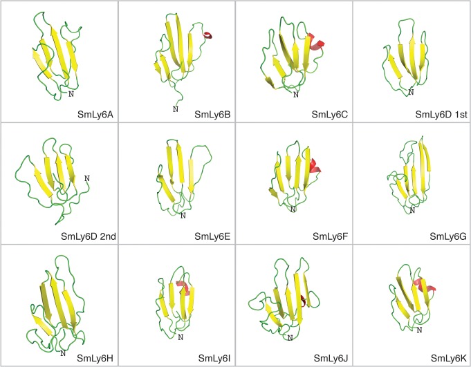
Fig 2. Structural models of SmLy6 sequences reveal common “three-fingered fold” tertiary structures.
SmLy6 tertiary structures were ab initio modelled using Rosetta software according to the Materials and Methods. From top to bottom, left to right ribbon representation of SmLy6A to SmLy6K where the two Ly6 domains of SmLy6D were modeled independently (SmLy6D 1st and SmLy6D 2nd). Beta strand, alpha helices and loops depicted in yellow, red and green respectively. Structural rendering generated using PyMOL.

