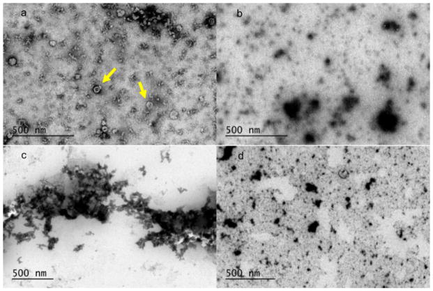
Fig. 7.
TEM images of uranyl acetate stained exosome fractions on a carbon grid. a Sample A, 12–20 min, numerous round dimple or cup-like structures [8] shown with arrow; b sample A, 24–32 min, irregularly shaped heterogeneous mixture of large and small particles; c sample B, 12–20 min, aggregates of many small particles; d sample B, 24–32 min, irregularly shaped heterogeneous mixture of large and small particles

