
FIG. 7.
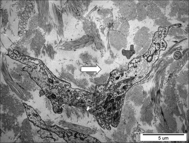
Electron microscopic picture of subcutaneous tissue in calf lymphedema stage III. Arrow at the site filled up with free fluid between collagen bundles. Excess stagnant fluid accumulated in the pericellular regions. X3000.
Official websites use .gov
A
.gov website belongs to an official
government organization in the United States.
Secure .gov websites use HTTPS
A lock (
) or https:// means you've safely
connected to the .gov website. Share sensitive
information only on official, secure websites.
Electron microscopic picture of subcutaneous tissue in calf lymphedema stage III. Arrow at the site filled up with free fluid between collagen bundles. Excess stagnant fluid accumulated in the pericellular regions. X3000.