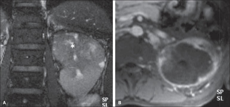
Figure 7.
Collecting duct RCC. A: MRI, coronal plane, T2-weighted image showing expansile, irregular lesion in the upper pole of left kidney, with heterogeneous signal intensity and predominance of hyposignal (asterisk). B: Contrast-enhanced MRI, axial, T1-weighted image. The lesion presents predominantly peripheral, heterogeneous signal intensity, considerably less intense than the renal cortex.

