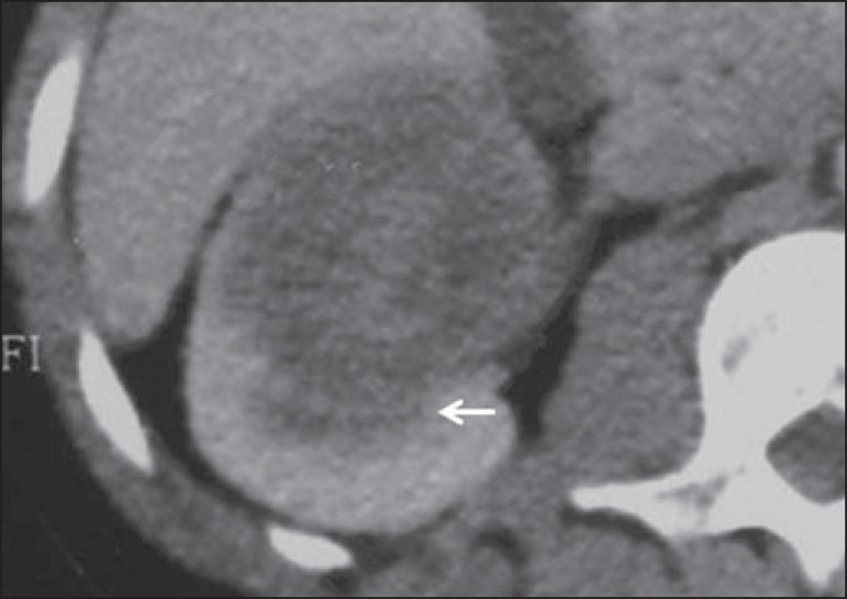
Figure 9.
Medullary RCC. Male, 25-year-old patient with sickle-cell disease. Contrast-enhanced CT image showing extensive, solid, hypovascular, predominantly medullary and slightly heterogeneous lesion in the right kidney. Observe the infiltrating feature of the lesion in the pyelocalyceal system (arrow) and in the proximal portion of the ureter.

