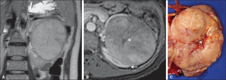
Figure 10.
Renal mucinous tubular and spindle cell carcinoma. Female, 57-year-old patient with hematuria. A: MRI, T2-weighted image showing expansile lesion with intermediate signal intensity, and (B) contrast-enhanced nephrographic phase showing hypovascular lesion – compare with the cortex (arrow). Observe the hyposignal of the scar (asterisk). Despite the large dimensions of the lesion, it is well delimited, with no infiltrating feature. C: Surgical specimen showing a circumscribed, yellowish lesion with central scar (asterisk).

