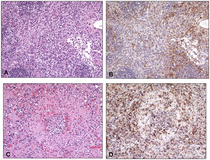
Fig 9. Histopathological and immunohistochemical findings in the inguinal lymph node and spleen.
Histopathological and immunohistochemical findings in the inguinal lymph node (A and B, Animal #13 (510 PFU group) and spleen (C and D, Animal #16 (48 PFU group). A) There is depletion of lymphocytes and replacement by inflammatory cells (macrophages and neutrophils) admixed with necrotic cells, necrotic debris, and fibrin. B) Poxviral antigen is present predominantly in mononuclear inflammatory cells. C) Diffuse depletion of white pulp with lymphocytolysis and necrosis. D) Antigen is abundant in cellular debris and multiple cell types (mononuclear inflammatory cells, endothelial cells, supporting stromal cells). Routine HE stain (A and C). Immunoperoxidase method with hematoxylin counterstain (B and D). All at 20X.

