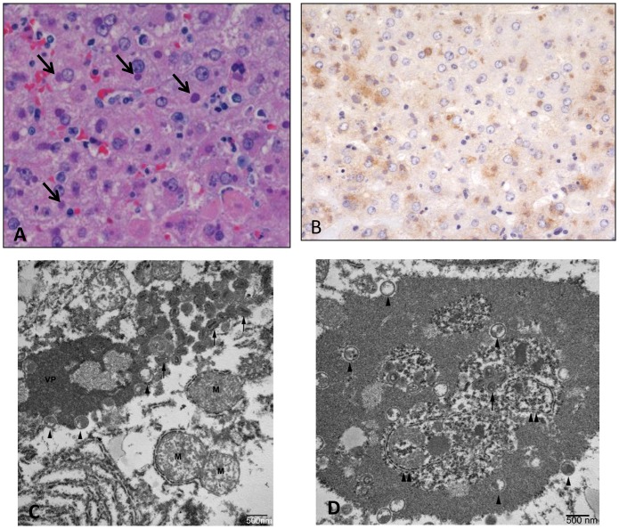
Fig 10. Histopathological, immunohistochemical, and electron microscopic findings in the liver.
Animal #3 (2.4 x 10⁷ PFU group). A) Hepatocellular degeneration and necrosis with prominent eosinophilic intracytoplasmic inclusions (arrows). HE. B) Immunohistochemistry demonstrates vaccinia viral antigen in the liver. Immunoperoxidase method with hematoxylin counterstain. C) Transmission electron micrograph of inclusion in hepatocyte. Note the varying stages of virion from immature (arrowheads) to mature (arrows). VP—viroplasm; M—mitochondira. D) Transmission electron micrograph of inclusion in hepatocyte containing endoplasmic reticulum (double arrowheads) and free ribosomes.

