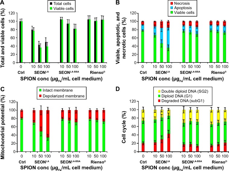
Figure 4.
Association of cellular uptake with toxicity of SPIONs. HUVECs were incubated for 48 hours with different amounts of SPIONs and cytotoxicity was analyzed by flow cytometry.
Notes: (A) Quantification of the absolute amount of total cell numbers and viable cells using MUSE. (B) Cell viability determined by Annexin V/propidium iodide staining. Percentages of necrotic (propidium iodide-positive), apoptotic (Annexin V-positive, propidium iodide-negative) and viable cells (Annexin V-negative, propidium iodide-negative) are shown. (C) Membrane potential analyzed by 1,1′,3,3,3′,3′-hexamethyl-indodicarbocyanine iodide [DiIC1(5)] staining. Graphs show cells with intact (DiI+) and depolarized (DiI−) membrane potential. (D) Cell cycle analysis by propidium iodide-Triton X staining. The cell status is expressed as the amount of apoptotic (degraded DNA, subG1), diploid (G1-phase) and double diploid (synthesis/G2 phase) cells. Data are presented as the mean ± standard error (n=3 with sample triplicates). The results were normalized to total cells (A) or to untreated control cells (B–D), set to 100%.
Abbreviations: SPION, superparamagnetic iron oxide nanoparticle; HUVECs, human umbilical vein endothelial cells; SEONLA, lauric acid-coated nanoparticles; SEONLA-BSA, lauric acid/albumin bovine serum hybrid-coated nanoparticles; ctrl, control.

