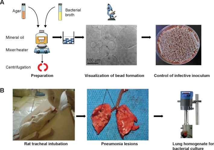
Figure 3.
Chronic pneumonia model using agar beads: main steps.
Notes: (A) Agar beads synthesis (top row of pictures). Broth containing bacteria and agar is added to mineral oil with continuous shaking and heating. The solution obtained is centrifuged to obtain beads whose size is measured and must be about 100 µM. The precise inoculum is assessed by serial dilutions method. (B) Model of rat pneumonia (bottom row of pictures). After inhaled anesthesia, the rat is suspended by the teeth and intubated into the trachea. Agar beads solution is injected into a tracheal catheter. After animal sacrifice, macroscopic aspects of lung lesions can be observed. Then, the lungs are homogenized for bacterial count.

