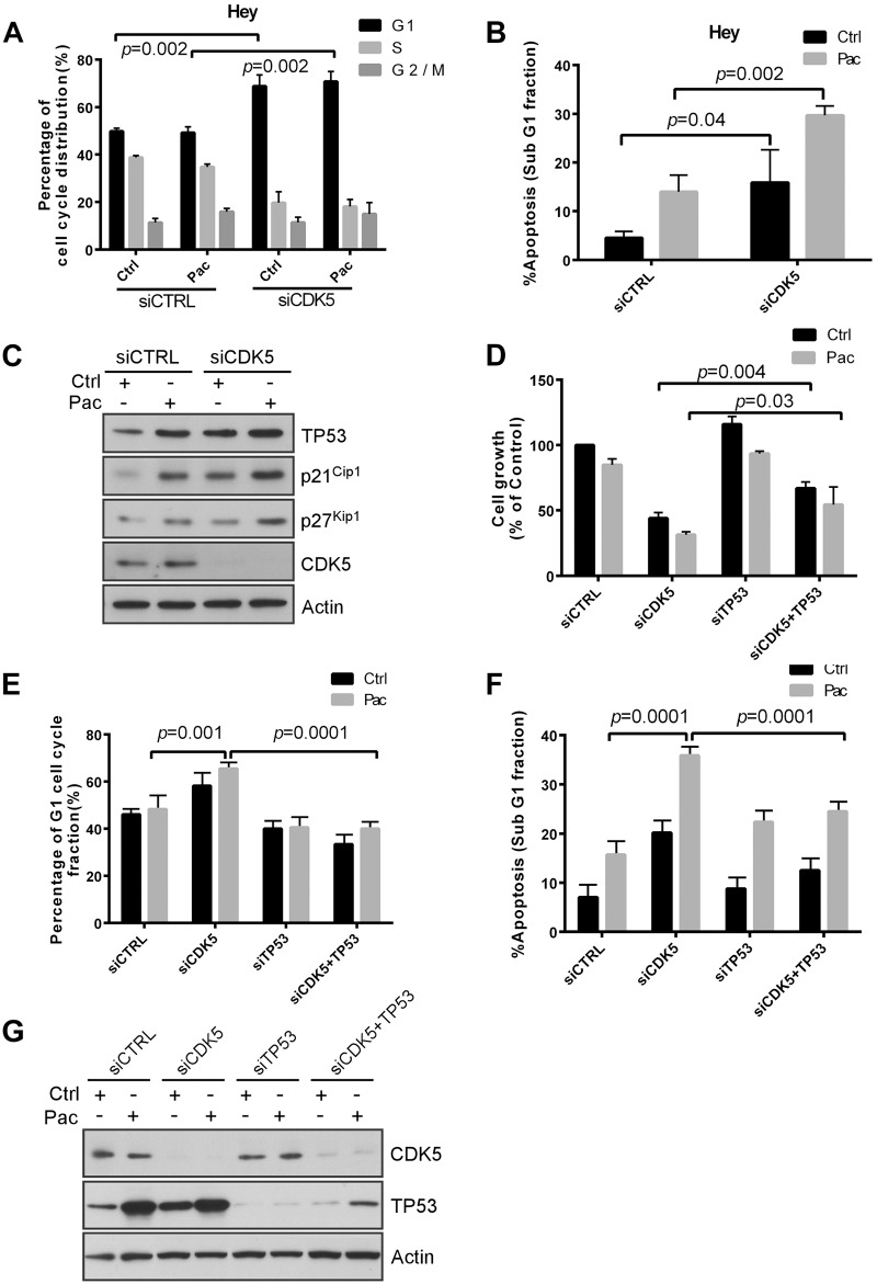
Fig 3. CDK5 knockdown induces p53-dependent growth inhibition, apoptosis and G1 arrest.
(A, B) Knockdown of p53 reduced CDK5 knockdown-induced apoptosis (A) and G1 arrest (B). HEY cells were reverse transfected with control siRNA or CDK5 siRNA for 24 h and treated with 5 nM paclitaxel (Pac) or diluent (Control) for an additional 48 h, and then cell cycle analyzed by flow cytometry. Data shown are mean values from three independent experiments. (C) CDK5 knockdown increased the expression of p21Cip1, p53, and p27Kip1. HEY cells were treated with control siRNA or CDK5 siRNA for 24 h and then with paclitaxel (3 nM) or diluent for 48 h. Immunoblot analysis was performed with antibodies against p21Cip1, p53, and p27Kip. (D) Knockdown of p53 expression reduced CDK5 siRNA-induced growth inhibition and reduced the enhancement of paclitaxel sensitivity. Cells were co-transfected with CDK5 siRNA and p53 siRNA for 24 h and treated with paclitaxel (Pac) or diluent. Proliferation of cells was measured with a crystal violet cell proliferation assay. (E, F) Knockdown of p53 reduced CDK5 knockdown-induced apoptosis (E) and G1 arrest (F). HEY cells were treated as in (A) and cell cycle analyzed by flow cytometry. Data shown are mean values from three independent experiments. (G) Western analysis confirmed increasing of TP53 expression by silencing CDK5. HEY cells were treated with control siRNA or CDK5 siRNA for 24 h and then with paclitaxel (3 nM) or diluent for 48 h. Cells lysates was subjected to immunoblot analysis with p53, CDK5 and actin antibody.

