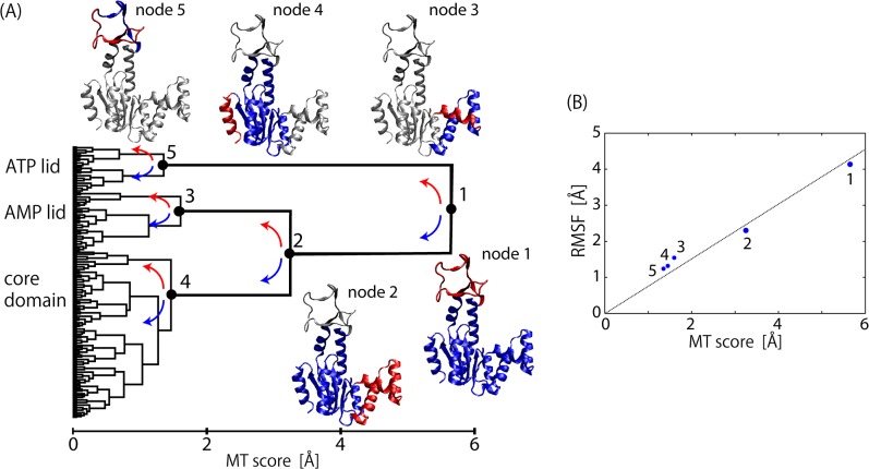
Fig 1. Motion tree for substrate-free ADK.
(A) Motion Tree constructed from 50-ns dynamics of substrate-free adenylate kinase. Five nodes are shown with corresponding parts of ADK structure in blue (larger domain) and red (smaller domain). (B) RMSF value for smaller (red) domain after fitting to corresponding larger domain is plotted at each node as a function of MT score. Dotted line is least square fit with zero at origin.

