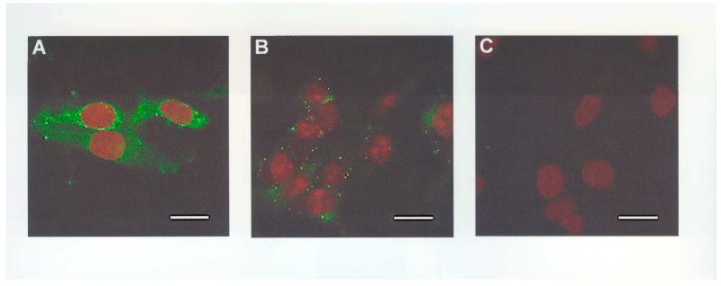
Figure 3.
Representative examples of the labelling of AβPP in MOG cells by 2B12 (A-C). (A) Punctuate labelling of MOG cytoplasm and perinuclear staining by 2B12 (5μg/ml) detected using a biotinylated anti-mouse antibody (1:270) and Avidin-FITC (1:600); (B) MOG incubated with 2B12 following pre-adsorption with Ka (10μg) and detected as above; (C) MOG incubated with the block solution alone, followed by detection as above. No labelling was detected in the absence of 2B12 or after pre-adsorption with Ka. Coverslips were mounted with VECTASHIELD mounting medium with propidium iodide to counter-stain the nuclei. Scale bar = 25μm, n=3.

