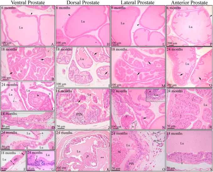
Fig 1. Histopathology of the prostatic complex of rats at different ages.
Ventral (A-G); dorsal (H-K); lateral (L-O) and anterior (P-S) prostate. (A, H, L and P) Prostates of young adult rats showed normal histology. (B, I, M and Q) Senile rats showed increased unfolding of the epithelium (arrows) in all prostatic lobes. (B) Luminal concretions (c). (C) Epithelial proliferation resulted in cribriform architecture with intraluminal growing (*) in the ventral prostate. (D) Mucinous metaplasia of the epithelium (bounded area). (E) Epithelial atrophy (a) and area of a cell with nuclear enlargement and prominence of nucleoli (n). (F and G) Hyperproliferative epithelium with characteristic foci of prostatic intraepithelial neoplasia (PIN). (G) Cells with evident nuclear enlargement (n) in the PIN area. (J) Detail of the prostatic intraepithelial proliferation observed in the bounded area of the dorsal prostate in panel I. (K) Epithelial proliferation with papillary growth. Thickening of the stroma (**) occurred around the lesion. (N) Lateral prostate with epithelial hyperplasia (arrow) as well as foci of inflammatory cells infiltrating the epithelium (bounded area), lumen (#) and stroma (i). (O) PIN area next to an inflammatory cell infiltrate containing plasma cells (Pl). Cells sloughed into the lumen (sc). (R) Intense unfolding and vacuolization in the epithelium of the anterior prostate (v). (S) PIN area in the anterior prostate with some nuclear atypia (n). Arrowhead = perialveolar smooth muscle cell; Pl = plasma cells; bv = blood vessel; c = luminal concretions; Lu = lumen. Stained with H&E.

