
Fig 2. Immunostaining for ERβ is affected in specific areas of alterations related to aging.
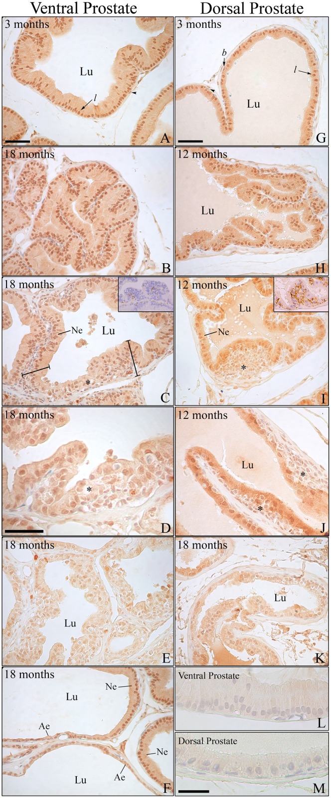
(A–F) Immunostaining for ERβ in the ventral and (G–K) dorsal prostate of rats at different ages. (A and G) Young adult animals showing intense positivity for the receptor in the epithelial luminal (l) and basal cells (b). Black arrowhead = positive perialveolar smooth muscle cells. (B and H) Senile rats presenting unfolding of the epithelium with normal ERβ expression. (C–D and I–J) Areas of intraepithelial proliferation (*) in the ventral and dorsal prostate, respectively, showing reduced ERβ staining compared with the normal epithelium (Ne). Insert in C and I: immunostaining for CK HMW in the ventral (C) and dorsal (I) prostate showing that the dorsal prostate presents a greater number of basal cells in intraepithelial proliferating areas. (E and K) Drastic reduction of ERβ immunostaining in areas of intense cellular atypia. (F) Transition from normal epithelium (Ne) showing standard ERβ positivity to atrophic epithelium (Ae) presenting a marked decrease in receptor staining. (L and M) Negative immunostaining controls for the ventral and dorsal prostate, respectively. Lu: lumen. Bar in A and G (= B, C, E, F, H, I, J, K) = 50 μm; bar in D = 40 μm; bar in M (= L) = 30 μm.