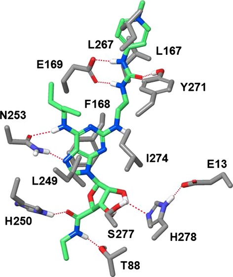
Figure 13.

Binding mode of compound 2 in the X-ray structure of human A2a in the agonistic conformation. Residues critical for potency and selectivity are indicated. The urea fragment of the C2-side chain is H-bonded to E-169 and Y-271. The terminal positively charged guanidinium moiety is exposed to the extracellular environment and squeezed between residues L167 and L267. H-bonds are shown by red dotted lines. Carbon atoms of compound 2 are shown in light green. Nonpolar hydrogens are not shown for clarity.
