
Figure 3.
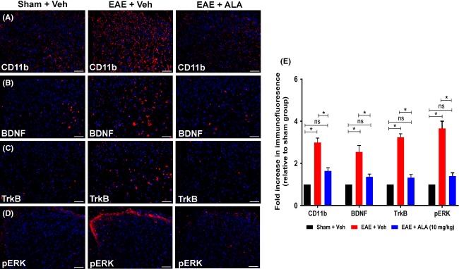
Immunohistochemical (IHC) analysis of CD11b, BDNF, TrkB, and pERK in the dorsal horn of lumbar (L4-L6) spinal cord sections from RR-EAE mice administered ALA at 10 mg kg−1 day−1 or vehicle, for 3 weeks relative to the corresponding data for vehicle-treated sham-mice. For vehicle-treated RR-EAE mice, there was a significant increase in the (A, E) extent of microglia/macrophage (CD11b) activation (approximately threefold; F(2,33) = 45.57; P < 0.05), as well as expression levels of (B, E) BDNF (∼2.5-fold; F(2,33) = 15.84; P < 0.05); (C, E) TrkB (∼3.2-fold; F(2,33) = 71.52; P < 0.05) and (D, E) pERK (∼3.6-fold; F(2,33) = 42.49; P < 0.05) c.f. the respective data for the lumbar spinal cord of vehicle-treated sham-mice. For RR-EAE mice treated with ALA (10 mg kg−1 day−1), lumbar spinal cord expression levels of CD11b, BDNF, TrkB, and pERK did not differ significantly (P > 0.05) from the respective data for lumbar spinal cord of vehicle-treated sham mice, *P < 0.05 (one-way ANOVA followed by Tukey’s multiple comparison test). Scale bars represent 50 μm. EAE, experimental autoimmune encephalomyelitis, Veh, vehicle; ALA, alpha lipoic acid; BDNF, brain-derived neurotrophic factor; TrkB, tyrosine kinase B; ANOVA, analysis of variance.