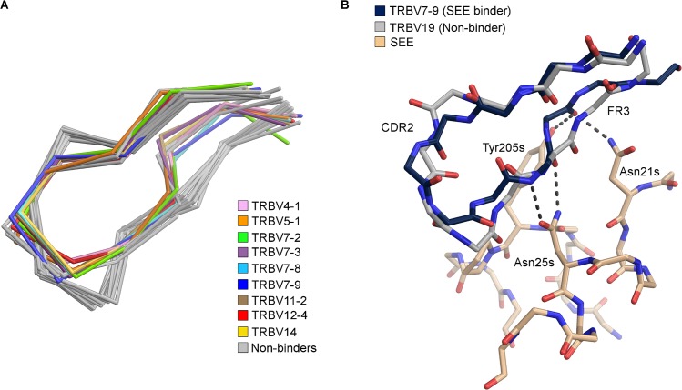
Fig 3. Comparison between structurally determined CDR2 loops.
(A) Close-up view of the CDR2β loop of 23 structurally determined TCRs. TRBV domains with coloured loops have been reported to bind SEE, while the non-binders are shown in grey; TRBV4-1 (pink), TRBV5-1 (orange), TRBV7-2 (green), TRBV7-3 (purple), TRBV7-8 (cyan), TRBV7-9 (blue), TRBV11-2 (brown), TRBV12-4 (red), and TRBV14 (yellow), (B) comparison between the SEE-TRBV7-9 structure in beige and blue, respectively, and TRBV19 in grey, which do not bind SEE. The hydrogen bond pattern to the backbone of CDR2 and the C” strand is shown with dotted lines.

