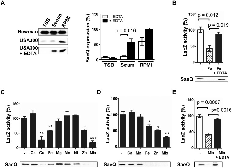
Fig 1. The SaeRS TCS can be repressed by Cu, Fe, and Zn.
(A) The effect of culture medium on the expression of SaeQ. S. aureus cells were grown to 0.5 OD600, and SaeQ protein was detected by Western blot analysis. Newman, S. aureus strain Newman; USA300, S. aureus strain USA300; + EDTA, addition of 1 mM EDTA. The quantification result of the Western blot is shown to the right. (B) The effect of iron on the P1 promoter activity (top) and SaeQ expression (bottom). P1 promoter activity was measured by LacZ assay for P1-lacZ fusion construct, while SaeQ was measured by Western blot analysis. -, no metal addition. (C) The effect of metal ions present in human blood on the P1 promoter activity (top) and SaeQ expression (bottom). S. aureus cells were grown in RPMI supplemented with 2 mM CaCl2, 20 μM CuSO4, 20 μM FeSO4, 500 μM MgSO4, 0.1 μM MnSO4, 0.04 μM NiSO4, or 20 μM ZnSO4 at 37°C for 16 h. Then the P1 promoter activity and SaeQ expression were measured by LacZ assay. Statistical comparison was made against the no metal (-) condition. (D) The effect of metal ions present in human neutrophil granules on the P1 promoter activity and SaeQ expression. Cells were grown in RPMI supplemented with 400 μM CaCl2, 130 μM FeSO4, 130 μM MnSO4, or 400 μM ZnSO4. All other conditions are the same as in (C). (E) The effect of metal chelation on the recovery of the P1 promoter activity (top) and SaeQ expression (bottom). S. aureus cells were grown for 16 h in RPMI supplemented with all metal ions (Mix) used in (D). The presented data represent three independent experiments. Error bars indicate standard error of the mean. Statistical significance was measured by unpaired, two-tailed t-test. *, p < 0.05; **, p < 0.01; ***, p < 0.001

