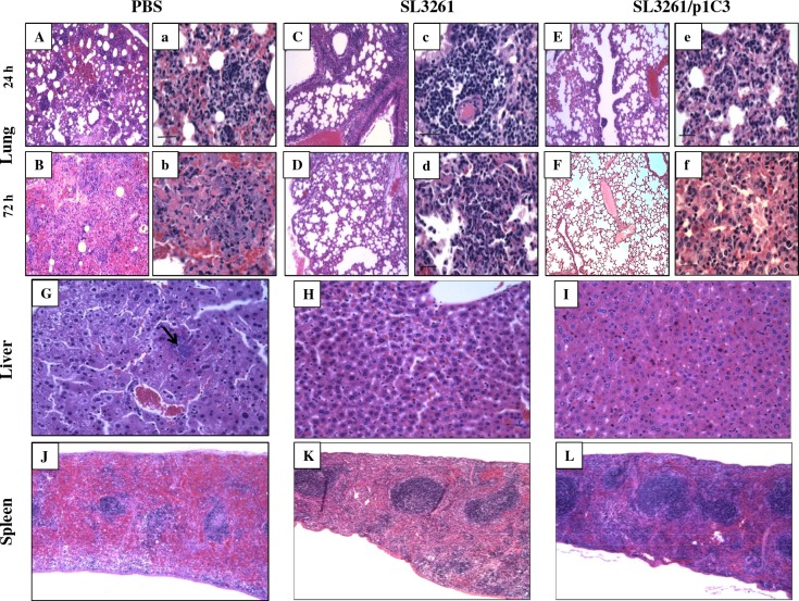
Fig 5. Histopathological changes in response to intranasal challenge with B. thailandensis E264.
Lungs of vaccinated BALB/c mice were isolated in their entirety at 24 (A-C) and 72 h (D-F) post-infection with 5 x 10⁶ CFU B. thailandensis (5 x LD50). The tracheas of each animal were exposed and inflated with 0.3 ml of 10% neutral-buffered formalin and immediately immersed in the same fixative. Livers (G-I) and spleens (K-L) were also used for histology; organs were collected under the same conditions. All samples were processed by standard paraffin embedding methods; sections were cut 2 mM thick and stained with haematoxylin-eosin (H & E). Preparation of tissue sections was performed by the University of Virginia Research Histology Core Facility. Tissue sections were examined by a veterinary pathologist who was blinded to animal group assignments. Sections A, B, C, D, E, and F are shown in magnification, 10x; representative sections a, b, c, d, e, and f are in magnification, 40x. Liver and spleen sections are shown in magnifications 10x and 5x, respectively.

