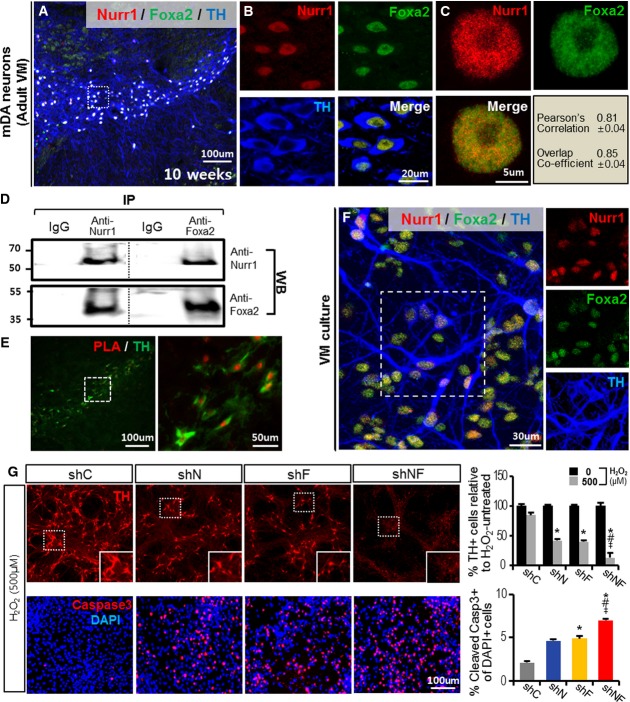
Nurr1 and Foxa2 interact physically and functionally to protect mDA neurons from toxic insult
A–C Co-localization of Nurr1 and Foxa2 proteins in mDA neurons of the adult mouse midbrain. Representative confocal sections of the ventral midbrain (A) stained with antibodies specific for TH, Nurr1, and Foxa2. The midbrains of mice at 10 weeks were cryosectioned, and stained images were taken using a confocal microscope with z-stacks through the section thickness (12 μm). Shown in (B) are individual and merged images of TH, Nurr1, and Foxa2 staining of the boxed area in (A) at higher magnification. Representative images of a single nucleus from a TH+ mDA neuron of an adult midbrain section (C) co-stained with Nurr1 (red) and Foxa2 (green). Co-localization of Nurr1 and Foxa2 in the merged images was assessed by Pearson's correlation and overlap coefficient values (n = 4, right/low quadrant in C).
D Immunoprecipitation (IP) assay for Nurr1 and Foxa2 protein binding. The ventral part of midbrains was dissected from 10-week-old mice, lysed, and subjected to IP. Nurr1/Foxa2 protein binding was detected by WB analysis using an anti-Foxa2 antibody in immunoprecipitates generated with anti-Nurr1 antibody (left), as well as by anti-Nurr1 WB assay in immunoprecipitates with anti-Foxa2 antibody (right).
E Physical interaction between Nurr1 and Foxa2 was further assessed by a proximity ligation assay (PLA). The SN area of a midbrain section (10 weeks old) was subjected to the PLA reaction and counterstained for TH. The boxed area in the left panel exhibiting physical Nurr1/Foxa2 interaction (red) in TH+ DA neurons (green) is enlarged in the right panel.
F, G Knockdown of Nurr1 and Foxa2 synergistically aggravates H2O2-induced cell death of mDA neurons in culture. Representative image for Nurr1- and Foxa2-co-expressing mDA neurons (F) used in the loss-of-function study. Shown in right panels are the individual Nurr1-, Foxa2-, and TH-stained cells of the boxed area. Nurr1- and Foxa2-co-expressing mDA neurons were formed after 9 days of differentiation in VM-NPC cultures in vitro and transduced with lentiviruses expressing shNurr1 (shN), shFoxa2 (shF), shN + shF (shNF), or shControl (shC). Three days later, the cultures were treated with H2O2 (500 μM) for 8 h and cells positive for TH (upper) and cleaved (activated) caspase-3 (lower) were counted the following day (G). Insets, high-power TH+ cell images of the boxed areas. Significantly different from the control (shC)*, shN#, shF‡ at P < 0.05, n = 5 culture wells in each group. P-values: 0.038 (shN*), 0.026 (shF*), 0.013 (shNF*), 0.036 (shNF#), and 0.042 (shNF‡) for the % TH+ cells; 0.043 (shF*), 0.037 (shNF*), 0.019 (shNF#), and 0.029 (shNF‡) for the percent cleaved caspase-3-positive cells; one-way ANOVA followed by Bonferroni post hoc test.

