Figure 2.
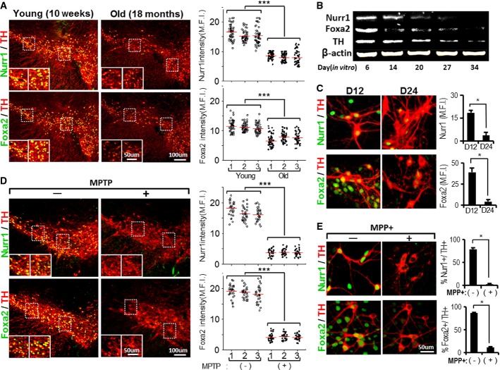
- A–C Nurr1 and Foxa2 protein levels in mDA neurons decrease in the midbrains of old mice in vivo (A) and in vitro after long-term culture (B and C). (A) Nurr1 and Foxa2 protein levels were compared in individual mDA neurons of the midbrains of young (10 weeks) and old mice (18 months) of the same mouse strain (C57BL/6, male). All the midbrain sections were immunofluorescently co-stained with Nurr1/TH (upper) and Foxa2/TH (lower) under identical conditions, and levels of Nurr1 and Foxa2 proteins were determined in individual TH+ mDA neurons by measuring mean fluorescence intensities (MFI) using LAS image analysis (Leica). Dots in the graphs represent the Nurr1 and Foxa2 MFI values of individual TH+ DA neurons in the SN of each animal. The average MFI values (indicated by horizontal lines) of three animals from each group were compared (***P = 5.25E-88 for Nurr1 intensity, 5.57E-40 for Foxa2 intensity, one-way ANOVA followed by Bonferroni post hoc test). Nurr1 and Foxa2 protein levels were also quantified in cultured mDA neurons over 6–34 days in vitro by Western blotting (B) and by immunocytochemical analysis (C). Significantly lower MFI values on day 24 of culture (D24) compared to D12 at *P = 0.027 (Nurr1), *P = 0.012 (Foxa2), n = 60–70 TH+ cells from two cultures in each group, unpaired Student's t-test.
- D, E Loss of Nurr1 and Foxa2 expression in mDA neurons after treatment with the neurotoxin MPTP (or MPP+). Mice (10 weeks old) were treated with MPTP for 5 days as described in Materials and Methods. Three days after the last MPTP injection, Nurr1 and Foxa2 protein levels in the TH+ mDA neurons of the MPTP-treated SN were compared with in the mDA neurons of untreated mice (D) (***P = 5.47E-103 for Nurr1 intensity, 1.53E-111 for Foxa2 intensity, one-way ANOVA followed by Bonferroni post hoc test.). The effects of neurotoxin treatment were also determined in mDA neuron cultures treated with MPP+ (250 μM, 8 h, E). *P = 0.015 (% Nurr1+/TH+ cells), *P = 0.018 (% Foxa2+/TH+ cells), unpaired Student's t-test.

