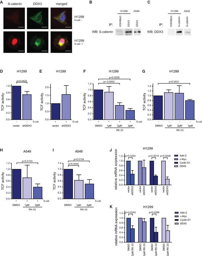
Figure 8.
- A β-catenin (red) and DDX3 (green) expression in H1299 cells. After overexpressing β-catenin, both DDX3 and β-catenin accumulate in the nucleus. Scale bar is 10 μm.
- B Immunoprecipitation with DDX3 or H3K4Me3 (control) and immunoblotted with β-catenin in A549 and H1299 cells. Outlined boxes indicate spliced lanes.
- C Immunoprecipitation with β-catenin or H3K4Me3 (control) and immunoblotted with DDX3 in A549 and H1299 cells. Outlined boxes indicate spliced lanes.
- D, E β-catenin/TCF4 activity was determined by the TOP/FOP reporter assay. Co-transfection with β-catenin is indicated below.
- F–I H1299 and A549 cells were treated with RK-33 (0, 1, 2, and 3 μM) and co-transfected with β-catenin in (F, H). Treatment with RK-33 decreased TCF4 activity in both cell lines.
- J, K Normalized mRNA expression of TCF4-regulated proteins (Axin-2, c-Myc, Cyclin D1) and DDX3 were measured by qRT–PCR in H1299 cells after knockdown of DDX3 (J) and treatment with RK-33 (K). All experiments were repeated three times.
Data information: Significance was assessed by two-sided, paired t-test. Error bars represent SD.
Source data are available online for this figure.

