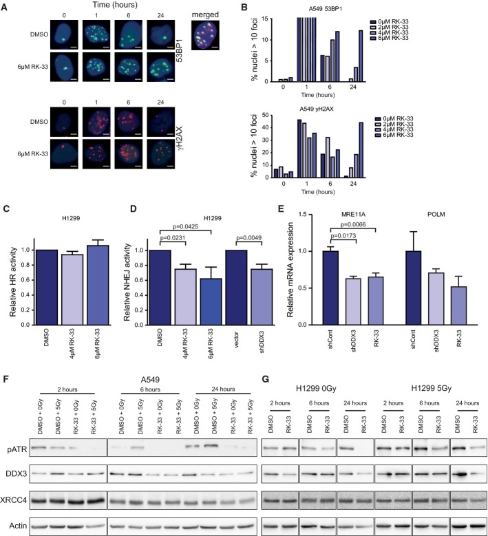
Figure 9.
- A Immunofluorescence images showing 53BP1 and γH2AX foci in A549 cells after 2-Gy radiation and A549 cells pre-treated with 6 μM RK-33, 12 h before radiation. Overlap of 53BP1 and γH2AX is seen in the merged picture of the co-immunofluorescence staining. Scale bar is 2 μm.
- B A549 cells were pre-treated with RK-33 and radiated with 2 Gy, and 53BP1 and γH2AX foci were counted as a measure of DNA damage. Cells with more than 10 foci 53BP1 or γH2AX were counted. More than 400 cells per sample were evaluated.
- C H1299 cells stably transfected with a homologous recombination (HR) reporter construct were treated with RK-33. Reporter constructs expressed GFP, which was quantified by flow cytometry. Experiments were repeated three times.
- D H1299 cells, containing a stable non-homologous end-joining (NHEJ) reporter construct, were treated with RK-33 and knockdown of DDX3. Reporter construct expressed GFP, which was quantified by flow cytometry. All experiments were repeated three times.
- E Microarray results from MDA-MB-231 cells treated with RK-33 and shDDX3 were validated by qRT–PCR using NHEJ Mechanisms of DSBs Repair PrimePCR plates (Bio-Rad) and performed in biological triplicates.
- F, G DNA repair-related proteins (ATR and XRCC4), DDX3, and actin were assessed by immunoblotting in A549 (F) and H1299 (G) cells. Cells were pretreated for 4 h with vehicle control or 6 μM RK-33 and then radiated with 0 or 5 Gy. Outlined boxes indicate spliced lanes.
Data information: Significance was assessed by two-sided, paired t-test. Error bars represent SD.
Source data are available online for this figure.

