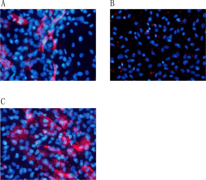
Fig 6. Alizarin Red S staining was positive for myofibroblasts.
Fibroblasts were incubated with control media for 4 days. Alizarin Red S staining of these cells was negative(Fig 5A). Calcification was not induced in the necrotic fibroblasts of mineralization group after incubation with mineralization media (DMEM containing 1% FBS, 50 ug/ml ascorbic acid, 5 mmol/L β-glycerophosphate) for 4 days. False positive was detected in fibroblasts by Alizarin Red S staining (Fig 5B).Calcification was detected in myofibroblasts of TGF-β1 (20 ng/ml) + mineralization group by Alizarin Red S staining (Fig 5C). Expression of α-actin in the myofibroblasts. Control group (Fig 6A), mineralization group (Fig 6B), TGF-β1 (20 ng/ml) + mineralization group (Fig 6C). Magnification *200

