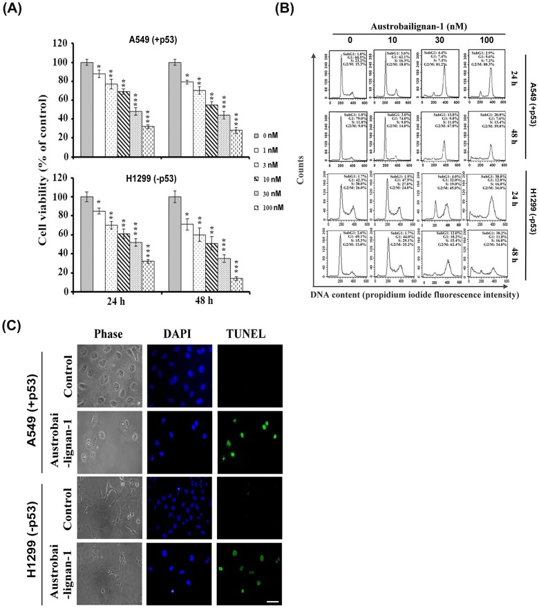
Fig 2. Austrobailignan-1 induced G2/M arrest and apoptosis.
(A) A549 and H1299 cells were treated with various doses (0, 1, 3, 10, 30 and 100 nM) of austrobailignan-1 for 24 and 48 h. Cell number was measured by a Trypan-blue dye exclusion method. Data are expressed as mean ± S.D. from 3 independent experiments. (*P <0.05,** P <0.01, *** P < 0.001 v.s. control). (B) Cells were treated with varied doses (0, 3, 10, 30 and 100 nM) of austrobailignan-1 for 24 and 48 h, and then stained with propidium iodide, and flow cytometry was performed to examine the cell cycle distribution. (C) Cells were treated without or with 100 nM austrobailignan-1 for 48 h,a TUNEL assay was then performed to detect apoptotic cells (green) and the nuclear DNA was stained with DAPI (blue). The stained cells were investigated by fluorescence microscopy. Magnification x 400; scale bar, 50 μm.

