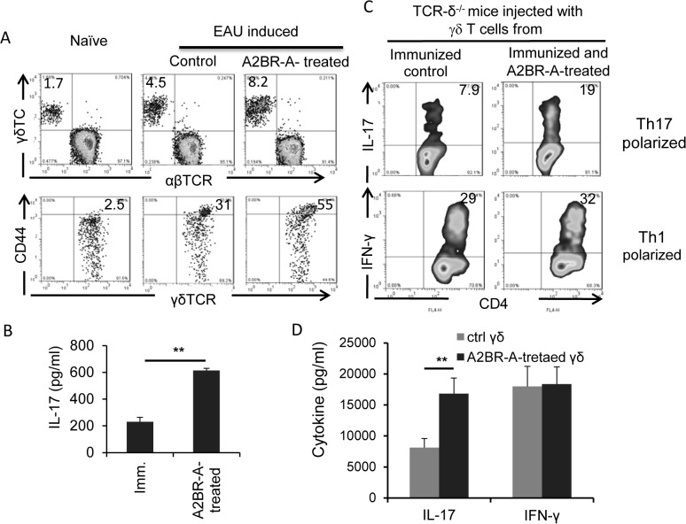
Fig 4. γδ T cells in A2BR agonist-treated mice were more activated in vivo and possesed increased Th17-promoting activity.
A) Proportional numbers of γδ T cells and their activation status were compared among γδ T cells in naive, immunized, and BAY60-6538-treated, immunized mice. Splenic T cells were isolated from pooled mice, stained with anti-αβ, anti-γδ or CD44 antibodies, and followed FACS analysis. B) Cytokine production of freshly isolated γδ T cells was examined after cultured for 48 h in vitro, in the absence of additional stimulation. C) In vivo reconstitution study showing that γδ T cells isolated from BAY60-6538-treated mice have increased Th17-promoting activity when injected to TCR-δ-/- mice. Groups of TCR-δ-/- mice (n = 6) immunized with the uveitogenic peptide (IRBP1-20/CFA) were injected i.p. at the day of immunization with 105 γδ T cells isolated from BAY60-6538-treated or untreated mice, before they were immunized with IRNP/CFA. 13 days post-immunization, splenic T cells were enriched and stimulated with immunizing peptide under Th1- and Th17-polarized conditions, and the activated T cells were stained for intracellular expression of IL-17 (top row) or IFN-g (bottom row). Results of one single experiment are shown, which were repeated for at least 3 times. D) 48-hour culture supernatants of the cells cultured in (C) were assessed for IL-17 and IFN-γ by ELISA. Data are from one single experiment, which are representative of three independent experiments **p<0.05.

