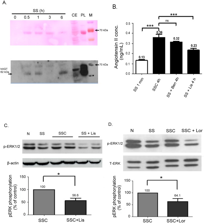
Fig 5. Detection of AGT and Ang II in a conditioned medium.
(A) Western blot analyses of AGT expression in an SS medium with different incubation times (0 minutes, 30 minutes, 1 h, 6 h). CE, cell extract from HUVECs. PL, plasma from blood sample. M, ladder marker. Upper panel shows rapid (5 minutes) staining of protein bands on a PVDF membrane with the Ponceau S stain. (B) The Ang II concentration detected using an ELISA assay was highly increased in the SS medium (conditioned for 4 h) compared with that in the ECs conditioned in the serum-free medium for 1 minute (***P < 0.001). Conditioning ECs with lisinopril in an SS medium, SS+Lis, suppressed the production of Ang II (***P < 0.001), but suppression was not found in SS+Ben. ns, no significance. Data are expressed as the mean ± SE and are representative of 3 experiments. (C) Western blot analyses of phosphorylated ERK1/2 expressions in VSMCs exposed to N, SS, SSC, and ACEi-SSC (SS+Lis) for 1 h. The activation of phospho-ERK1/2 was suppressed in the ACEi-SSC group. (D) The activation of phospho-ERK1/2 in SSC was suppressed by 10 μM losartan.

