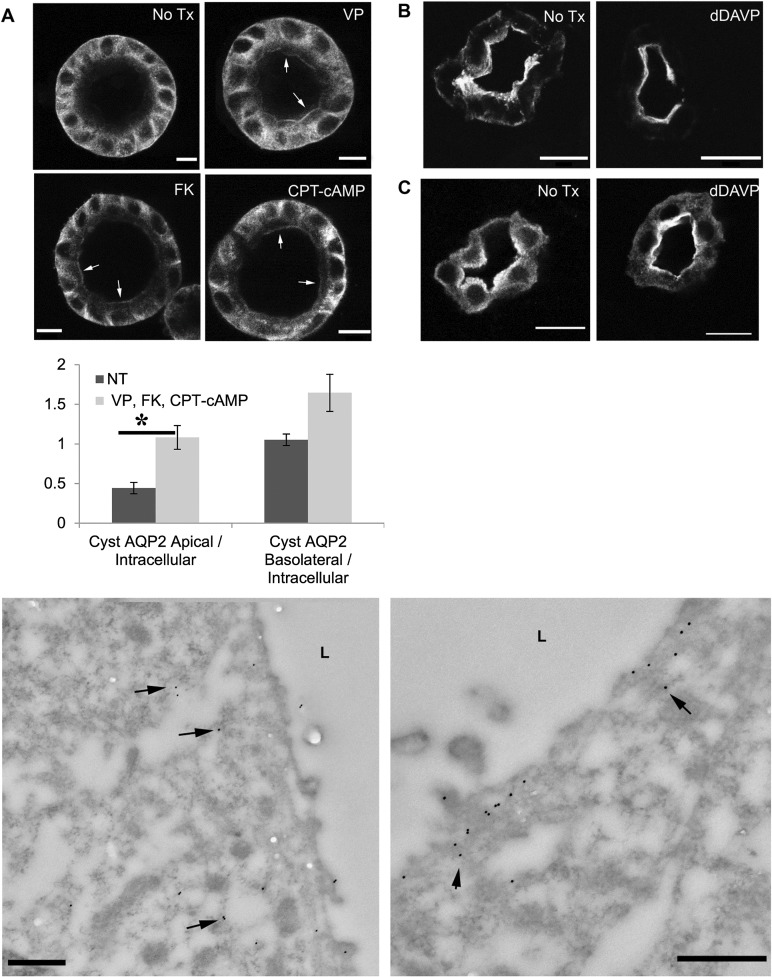
Fig 3. Regulated trafficking of AQP2 is intact in MDCK cysts.
AQP2 trafficking in MDCK-AQP2 cysts is intact and staining patterns for total AQP2 are comparable to those observed in Brattleboro, and normal rat kidney. (A) MDCK-AQP2 cysts were incubated in serum free medium for 120 minutes. Addition of AVP, FK, or CPT-cAMP to the medium for 40 minutes resulting in apical membrane accumulation of AQP2 (arrows). Bar = 10 μm. (D) Asterisk denotes that significantly (P < = 0.05) more apical, but not basolateral staining of AQP2, relative to intracellular AQP2, was observed following stimulation with AVP, FK or CPT-cAMP. N = 5 cysts (NT, non-treated), N = 12 cysts (AVP/FK/CPT-cAMP stimulated). The data for AVP, FK and cAMP-treated cysts were pooled together because we saw no statistically significant difference in Apical/Internal total AQP2 or Basolateral/Internal total AQP2 between the treatment modalities. (B) In the Brattleboro rat kidney AQP2 was located mainly in the subapical region while apical membrane accumulation of AQP2 was seen after treatment with dDAVP for 3 days. Bar = 10 μm. (C) Similarly, in a tissue slice culture from normal rat kidney, incubation in medium without VP resulted in AQP2 in the cytosol and subapical region. dDAVP treatment for 20 minutes resulted in AQP2 translocation to the apical membrane, with AQP2 still detectable in the cytosol. (E) In transmission electron micrographs, AQP2 in the MDCK-AQP2 cyst is labeled with 15nm gold particles. AQP2 gold particles distributed diffusely throughout the cytosol under baseline, non-stimulated conditions (left panel) while AQP2 accumulated on the apical membrane after VP stimulation (right panel), but not on the basolateral membrane (S2 Fig). Bars = 500 nm

