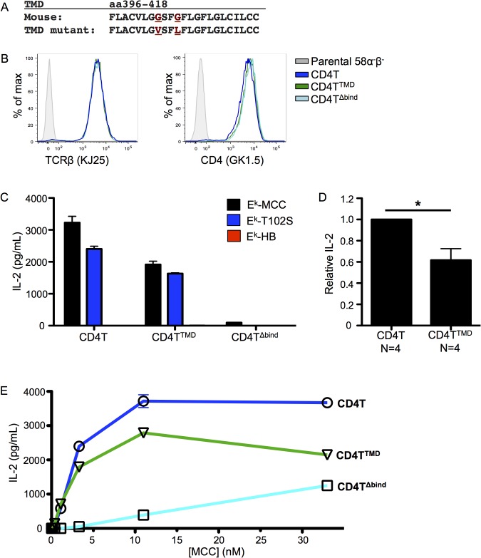
Fig 3. Mutating the CD4 transmembrane domain GGxxG motif impairs T cell activation.
(A) Alignment of the wild-type CD4 TMD with the CD4 TMD mutant (G403V/G406L). Mutated residues are highlighted in red. Bulky side chains were introduced to disrupt the glycine patch. (B) 58α-β- T cell hybridomas were retrovirally transduced with the 5c.c7 TCR and either CD4T, CD4TTMD or CD4TΔbind, which is mutated in the region known to bind MHC class II. Surface expression of TCR and CD4 were assessed by flow cytometry as labeled. (C) IL-2 secretion from 58α-β- T cell hybridomas after 16 hours of co-culture with M12 cells expressing agonist (MCC), weak agonist (T102S), or null (HB) peptide tethered to I-Ek. Data are representative of four independent experiments with independently generated cell lines. (D) IL-2 secretion from four independently generated CD4TTMD 58α-β- T cell hybridomas after 16 hours of co-culture with MCC:I-Ek+ M12 cells normalized to matched CD4T controls within the same experiment to determine relative IL-2 concentration. (E) IL-2 secretion from 58α-β- T cell hybridomas after 16 hours of co-culture with Chinese hamster ovary (CHO) cells ectopically expressing I-Ek (CHO Ek) pulsed with MCC peptide at the indicated concentrations. Data are representative of three independent experiments with independently generated cell lines. (*p<0.05; Mann-Whitney).

