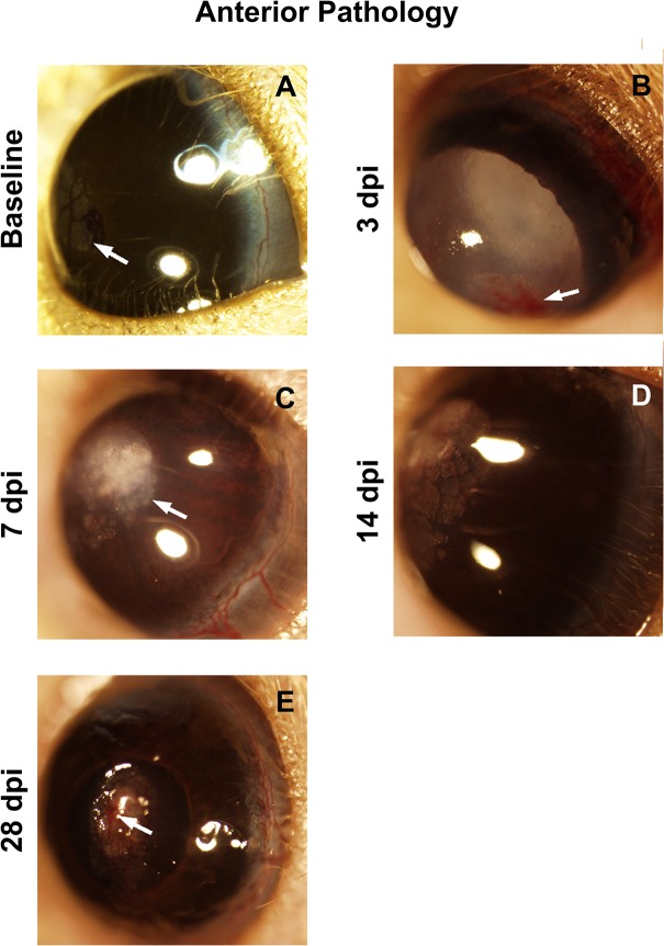
Fig 1. The ocular surface of the D2 eye is injured.
(A) Calcium deposits (arrow) are common in the normal D2 eye. (B) Hyphema (arrow), corneal edema and a cataract at 3 dpi. (C) Corneal edema and calcium deposits (arrow) at 7 dpi. (D) Corneal edema, corneal neovascularization and calcium deposits at 14 dpi. (E) A corneal scar and corneal neovascularization (arrow) at 28 dpi.

