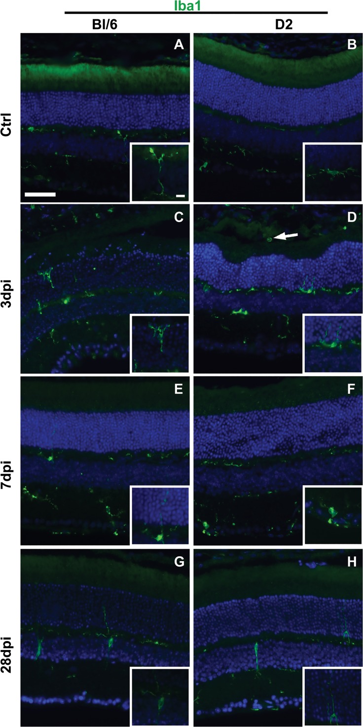
Fig 9. Reactive microglia are present in Bl/6 and D2 retinas after injury.
Low magnification epifluorescence micrographs and high magnification micrographs (insets) of control (A-B), 3 dpi (C-D), 7 dpi (E-F) and 28 dpi (G-H) retinas immunolabeled with Iba1 (green) and DAPI (blue). The scale bar for the low magnification micrographs is 50μm. The scale bar for the inserts is 10μm.

