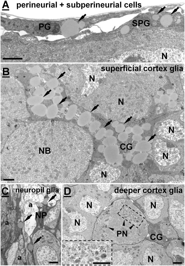
Fig 3. Representative electron micrographs of particular glial cell types.
LDs are marked by arrows. (A) Perineurial (PG) and subperineurial cells (SPG) located on the surface of the brain. (B) A superficial glial cell located in the outer layer of the brain cortex, containing high amount of LDs. (C) A neuropil glia (NP) located at the cortex-neuropil boundary ensheating axons. (D) A deeper cortex glia (CG) found close to the cortex-neuropil boundary encapsulating a large peptiderg neuron (PN) and several other neurons (N) with its processes. No LDs seen in the cytoplasm of such a cortex glia. Note the presence of large clusters of neurosecretory vesicles (arrowheads) in the cytoplasm of PN. Scalebar: 2μm.

